Optical instruments
Human eye
The basic structure of the human eye is shown in the figure below.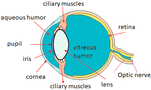
Cornea is the transparent front part of the eye, its refractive index is 1.376.
Vitreous humor is the interior of the eye, which is filled with a transparent gel-like substance. Its index of refraction is 1.337.
Lens: The lens has a varying refractive index, from 1.386 to 1.406.
Aqueous humor is a watery fluid between the cornea and lens. Its refractive index is 1.336.
Iris is a diaphragm, and a colored part of the eye. It adjusts automatically to control the amount of light entering the eye.
Pupil is a hole in the iris through which light passes. It appears black.
Retina is a curved light sensitive rear surface of the eye. It consists of a complex array of nerves and receptors known as rods and cones.
Image reconstruction
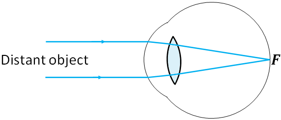
Light falls on the eye is refracted at the front surface of the cornea. Then, the eye lens bends (refracts) the light rays and are focused on the retina. Most of the bending of light rays is done at the cornea. Bending by the lens is small as compared to that by the cornea. At the retina, the light energy is converted into electrical signals. These electric signals are then transmitted to the brain by the optical nerves. The reconstruction of the image is done at the brain.Focusing and accommodation
Based on the object's distance from the eye, the focal length of the eye lens changes. The focal length is changed by changing the curvature of the lens, which is done by the ciliary muscles.When focusing on a distant object, the ciliary muscles of the eye are relaxed and the lens becomes thin, which shortens focal length and the parallel rays are focused on the retina (the focal point, F).
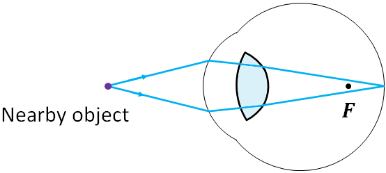
To focus on a nearby object, the ciliary muscles contract, which makes the center of the lens thicker and the focal length of the lens shortens. So, the image of the objects is focused on the retina. These focusing adjustments of an eye is called accommodation.
Near point and far point
The closest distance at which an eye can focus is called the near point of the eye. For young adults, the near point is 25 cm. Near point could be as close as 10 cm for younger children. As people grow older, near point increases and the ability to accommodate is reduced. We take 25 cm as the near point for a normal human eye.Far point is the farthest distance at which an object can be seen clearly by an eye. For a normal eye, the far point is greater than 6 m. When doing problems, we take the far point as infinity for a normal eye. This is because, when you use the lens equation, there will be not much difference if you use 6 m or infinity as the far point.
Defects of eye
Normal eyes focus the image of an object on the retina. But due to various eye defects, in some eyes, the image is focused in front or behind the retina.Nearsightedness or myopia
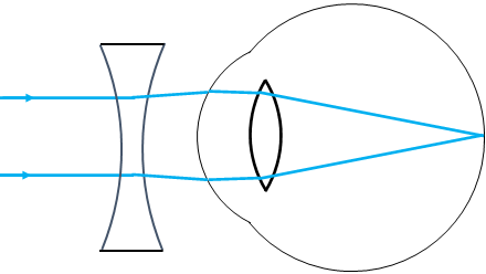
Some eyes can focus only on nearby objects and the distant objects cannot be seen clearly. This defect of an eye is called nearsightedness or myopia. Nearsightedness is caused by an eyeball that is too long. In a nearsighted eye, the images of distant objects are focused in front of the retina.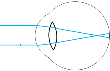
To correct nearsightedness, a diverging lens is used. A diverging lens allows the rays to be focused at the retina.
Farsightedness or hyperopia
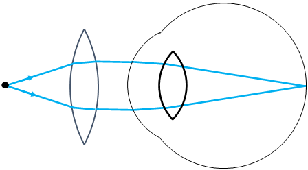
Another defect of an eye is the farsightedness or hyperopia. This defect is caused by an eyeball that is too short. The eye cannot focus on nearby objects but distant objects are seen clearly. The near point of a farsightedness eye is greater than 25 cm. In a farsighted eye, the images of nearby objects are focused behind the retina.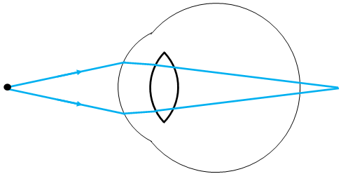
To correct farsightedness, a converging lens is used.
Contact lenses
Contact lenses are placed directly on the cornea to correct eye defects.Magnifying glasses
A magnifying glass is a converging lens that is used to produce a magnified (enlarged) image of an object. When we look at an object, the image size of the object that we see depends on the angle subtended by the object at our eyes. To see an enlarged image, the angle needs to be greater. To increase the angle, the object needs to be brought closer to our eyes. But we cannot bring the object closer than to the near point as we cannot see clearly anything closer than the near point.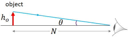
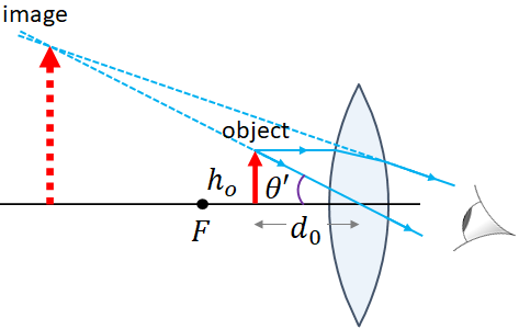
Angular magnification
The angular magnification or magnifying power, $M$ of a magnifying glass (lens) is defined as
$M=\dfrac{\theta'}{\theta}$.
where $\theta$ is the angle subtended by the object at the unaided eye (first figure), and $\theta'$ is the angle subtended by the object at the lens (second figure).From the figure, we can write
$\tan \theta =\dfrac{h_o}{N}$
where $h_o$ is the height of the object.Assuming the angle, $\theta$ is small, we can write
$\theta \approx \tan\theta = \dfrac{h_o}{N}$
From the second figure, we can write,
$\tan \theta' =\dfrac{h_o}{d_o}$
For small $\theta'$, we can write
$\theta' =\dfrac{h_o}{d_o}$
Substituting, $\theta$ and $\theta'$ in the angular magnification equation,
$M=\dfrac{N}{d_o}$.
Object at the focal point of the lens
Our eyes are relaxed when it receives parallel rays. So, when we place a magnifying glass, we place it at the focal point of the lens that makes the rays from the object parallel. When the object is at the focal point, $d_o=f$, where $f$ is the focal length of the magnifying glass. So, the angular magnification is
$M=\dfrac{N}{f}$.
Image at the near point
We can increase the angular magnification by one unit higher if we adjust the lens so that the image of the object is at the near point of the eye as follows.If the image is at the near point, then $d_i=-N$, where $N$ is the near point.
From the lens equation, we have,
$\dfrac{1}{d_o}=\dfrac{1}{f}-\dfrac{1}{d_i}$
Substituting $d_i=-N$,
$\dfrac{1}{d_o}=\dfrac{1}{f}+\dfrac{1}{N}$
Multiplying $N$ on both sides,
$\dfrac{N}{d_o}=\dfrac{N}{f}+1$
The left hand side is $M$, therefore,
$M=\dfrac{N}{f}+1$
Aberrations of lenses and mirrors
Spherical aberration
You saw that in lenses and in mirrors, parallel rays are focused at the focal point of the lens or the mirror. But in reality, not all parallel rays are focused at one focal point. The parallel rays near the principle axis of a lens (or mirror) are focused at different point than those near the edges of the lens (or mirror). This is called spherical aberration. It causes blurred images.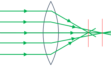
The spherical aberration can be minimized by using only the central part of the lens and avoid using the edges. Using aspherical lens or combination of lenses also reduces the spherical aberration.
Chromatic aberration
Bending (refraction) of light in a lens depends on the refractive index of the lens. The refractive index of a medium such as a lens depends on the wavelength of the light. White light has all the colors (i.e., many wavelengths), so, when white light is incident on a glass lens, different colors are focused at different points as blue light bends more due to its higher refractive index than red. This is called chromatic aberration. Chromatic aberration creates color distortion in images.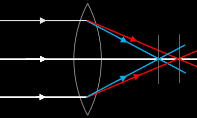
Chromatic abberration can be reduced by making lenses with glass materials of low dispersion, and also with other optical techniques.
Limits of resolution- circular aperture
Resolution of a lens is its ability to produce distinct images of two objects that are very close to each other. The closer the two images of the object and still be seen as distinct instead of overlapping blobs, the higher the resolution. The two factors that limit the resolution of a lens are aberration and diffraction. We can eliminate the aberration but we cannot avoid diffraction.Image and diffraction
When light from a point source passes through a circular aperture (that is a spherical lens or a pin hole), you will see a bright central spot surrounded by fainted rings due to diffraction. The pattern is called diffraction pattern or Airy pattern and the central bright spot is called the diffraction spot or Airy disk.
The intensity of light is maximum at the center of the disk. If you draw a graph of the intensity, $I$ at different points of the diffraction pattern versus the angle, $\theta$ of the point measured from the direction of propagation of the light, you will get the following graph.
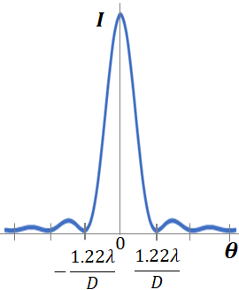
It has been found that the center spot covers the angle from $-\dfrac{1.22\lambda}{D}$ and $\dfrac{1.22\lambda}{D}$, where $\lambda$ is the wavelength of the light and $D$ is the diameter of the lens or the hole.
So, if $\theta_w$ is angular width of the airy disk, then
$\theta_w = \dfrac{2.44 \lambda}{D}$
Rayleigh criterion-resolution
If we have two point light sources, then we will see two Airy patterns. Note that the light sources can be any two distant objects, or two points on an object. If the two sources are close to each other, then the Airy patterns of the two sources overlap. But how close the two images can be and still we can tell that the images represents two sources instead of one?.Lord Rayleigh developed a criterion for the resolvable angular separation between two point objects. According to the Rayleigh criterion, two images are just resolvable when the center of the diffraction disk of one image is directly over the first minimum in the diffraction pattern of the other as shown in the figure below. The first minimum is the dark space between the center disk and the first ring.
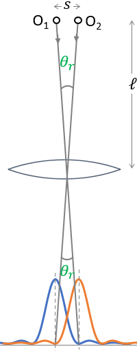
In the figure, there are two objects O1 and O2, being imaged by a lens. According to the Rayleigh criterion, at the minimum, the images of the two objects should overlap as in the bottom of the figure to resolve. And the angle, $\theta_r$ is the angle between the center of the Airy disk and the first minimum. Comparing this figure with the graph of the intensity versus angle graph of the image of a point source, we have
$\theta_r=\dfrac{1.22\lambda}{D}$
This angle is the minimum angular separation between the two objects for them to be resolved, and is called the angular resolution.We can find the spatial resolution, $s$ that is how close two objects can be so that we can resolve them with a lens, from the angular resolution. If $\ell$ is the distance between the lens and the object, then the resolvable distance between the two objects is
$s=\ell\,\theta_r$
Resolution of the human eye
Our eyes are more sensitive to the color of light of wavelength, $550 \,nm$. Typical angular resolution of a human eye is$\theta = 5\times 10^{-5} rad.$
For objects at the near point, eye can resolve objects that are about $0.1 \,mm$ apart.